Photo Galleries: Three Dimensional Lepidoptera Genitalia
Photo Gallery: Rhopalocera
Male genitalia images for dissection
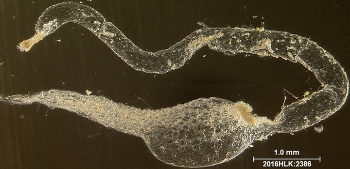
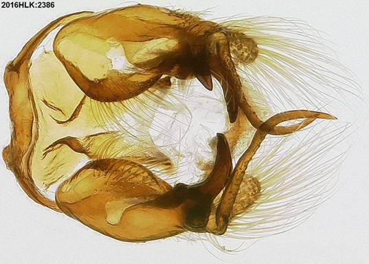
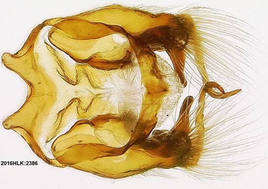
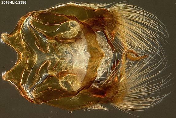
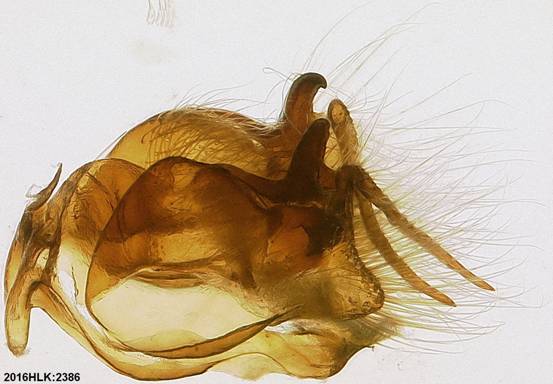
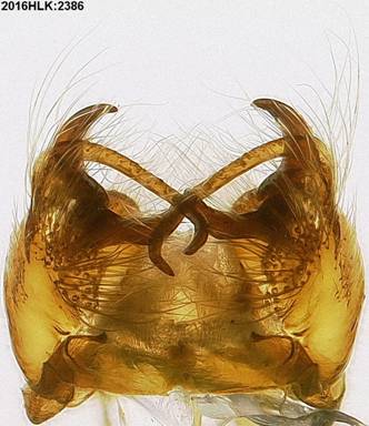
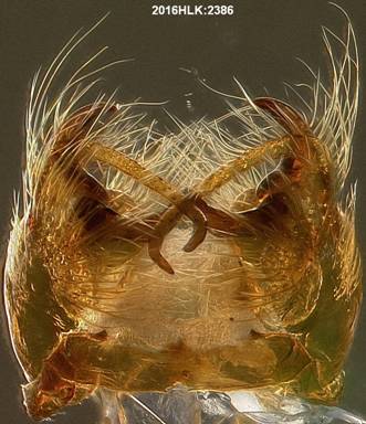
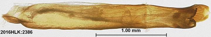
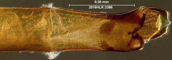
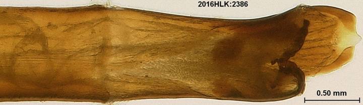
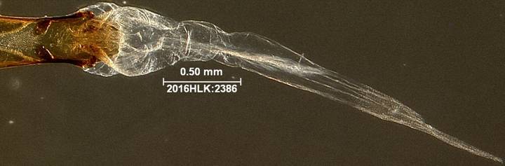
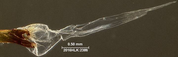
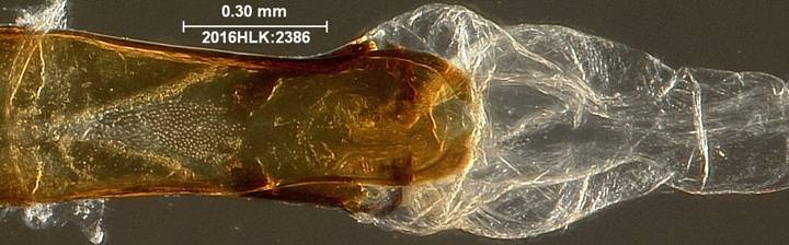
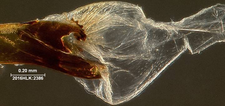
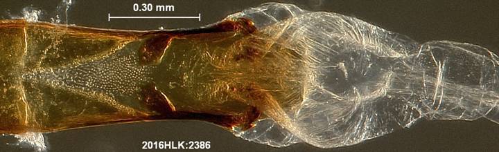
voucher specimen 2016HLK:2386 of Chlosyne janais (Nymphalidae: Nymphalinae: Melitaeini)
Genitalia were dissected and images taken by Hugo L. Kons Jr. with the GT Vision imaging system at the former American Entomological Institute. All structures are the natural three dimensional shape (not slide mounted).
For higher quality images but slower downloads, view in microsoft internet explorer. For lower quality images but faster downloads, view in Mozilla Firefox.
Adult
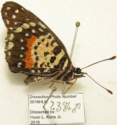
TX Medina County: Hondo Creek, 10 June 2008, Hugo Kons Jr.
Capsule
(ventral)
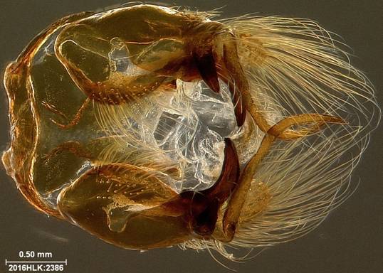
Capsule
(dorsal)
Capsule
(lateral with hairs and scales)
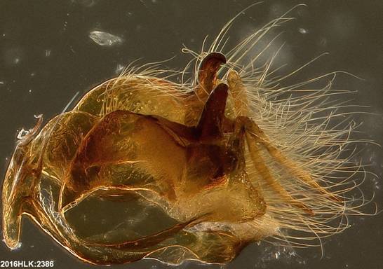
Capsule
(posterior)
Phallus
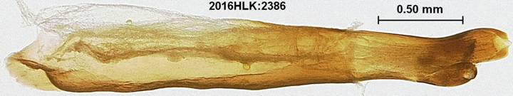
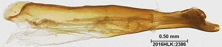
Phallus
apex with vesica and ductus
ejaculatorius everted
Vesica
(with hood above)
Vesica
(lateral, with hood on bottom)
Vesica
(with hood below)
Rectum
and Intestine